Ureaplasma urealyticum naturally colonizes the lower urogenital tract in up to 40% to 80% of healthy, sexually active adults. Its simple presence rarely signifies overt disease.
Standard methods for diagnosis, such as routine nucleic acid amplification tests (NAAT/PCR) do not provide a distinction between harmless colonization and active tissue infection.
Similarly, serological testing (IgG and IgM antibodies) fails to bridge this diagnostic gap, as circulating titers reflect past exposure, baseline mucosal colonization, or non-specific humoral activation rather than localized mucosal invasion.
This diagnostic ambiguity creates the risk of over-prescribing broad-spectrum antimicrobials for benign colonization, which can accelerate global antimicrobial resistance (AMR) in Mollicutes.
Under-treating genuine tissue invasion can threaten reproductive health, gestational outcomes, and neonatal viability.
Quantitative Thresholds (qPCR & Bacterial Load)
Load-Dependent Pathogenicity
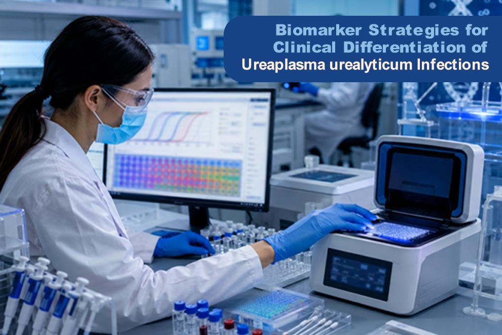
Researchers use real-time PCR (qPCR) to evaluate load-dependent pathogenicity to overcome the limitations of qualitative detection. Ureaplasma urealyticum is a pathogen that has the capacity to induce tissue inflammation, which correlates directly with high bacterial density.
Ureaplasma urealyticum typically persists as a benign commensal below a certain genome copy threshold. However, rapid proliferation triggers host immune activation and localized tissue damage.
Establishing Threshold Cutoffs
Researchers distinguish active infection from normal colonization using a threshold of 10,000 DNA copies per milliliter. When the concentration of Ureaplasma is higher than this limit, it may indicate conditions such as:
- Non-gonococcal urethritis (NGU) in men
- Chorioamnionitis, premature rupture of membranes (PROM), and preterm labor in pregnant women
Technical Challenges
Sample Matrix & Dilution Differences
The concentration of bacteria varies depending on whether a sample comes from:
- Diluted urine
- A mucosal swab
- or amniotic fluid
DNA Copies vs. Live Bacteria
Quantitative PCR detects genetic material from both living and dead bacteria, which can artificially inflate estimated bacterial loads.
Lack of an International Reference Standard
Without a universal calibration standard, quantitative results cannot be reliably compared across different research laboratories.
Host Immune Biomarkers & Local Inflammatory Cascades
Mucosal Immunity vs. Systemic Antibodies
Evaluating local host immune responses provides a far more accurate assessment of disease activity than circulating serum antibodies. These bacteria mostly colonize the mucosal lining of the host. As a result, systemic antibody levels (IgG/IgM) often reflect historical exposure or benign colonization.
Targeted diagnostic tools, such as a UPP2 ELISA kit, can be used to evaluate specific host tissue proteins and immune response indicators in local samples. When bacteria invade tissue, host cells release chemical signals (cytokines), which are measured to reveal an active, harmful infection.
Key Pro-inflammatory Markers
When U. urealyticum transitions from a commensal state to an active pathogen, host cells release specific pro-inflammatory cytokines:
Interleukins (IL-6, IL-8, IL-1β)
Ureaplasma urealyticum lacks a rigid cell wall. As a result, it presents membrane lipopeptides directly to host cells, which trigger the secretion of IL-6, IL-8, and IL-1β.
Interleukins trigger rapid inflammatory responses by recruiting white blood cells.
Tumor Necrosis Factor-alpha (TNF-α)
Elevated TNF-α and interleukins work in synergy to activate matrix metalloproteinases (MMPs). Localized MMP secretion breaks down collagen in fetal membranes, driving cervical ripening, premature rupture of membranes (PROM), and preterm labor.
Toll-Like Receptor Pathways
Surface lipoproteins on U. urealyticum bind directly to TLR-2 and TLR-4. As a result, white blood cells are recruited to the area, which causes inflammation. This inflammation is tracked to distinguish between colonization and infection.
Bacterial Virulence Markers & Phase Variation
Multiple Banded Antigen (MBA)
The multiple banded antigen (MBA) is the predominant surface-exposed lipoprotein and key immunogen of Ureaplasma urealyticum. The gene encoding MBA contains a hypervariable region subject to high-frequency phase variation and size variation. By rapidly altering the length and expression of its MBA surface proteins during infection, U. urealyticum continuously alters its antigenic profile. This high-frequency phase variation allows the bacterium to evade host antibody responses, persist on mucosal surfaces, and establish chronic, invasive infections.
Urease Biomineralization & Metabolic Biomarkers
U. urealyticum relies on urea as a primary metabolic energy source. It utilizes a surface-associated urease enzyme complex to hydrolyze urea into ammonia and carbon dioxide. Active urea hydrolysis can cause localized hyperammonemia, which directly disrupts the integrity of host mucosal epithelial layers through cytoplasmic alkalinization and cellular toxicity.
This metabolic activity yields valuable diagnostic biomarkers of tissue damage, including notable local pH shifts toward alkalinity and the generation of reactive oxygen species (ROS) that exacerbate cell damage.
Genomic Subtyping
Genomic subtyping provides crucial insight into invasive risk. Molecular assays can now distinguish Ureaplasma parvum (Biovar 1) from Ureaplasma urealyticum (Biovar 2). Advanced testing goes a step further by identifying specific serovars linked to severe disease.
Specific serovars express distinct virulence factors that directly drive invasive potential. These high-risk strains are strongly linked to ascending genital tract infections, persistent chorioamnionitis, and adverse neonatal outcomes.
Conclusion
Qualitative PCR is not reliable enough to differentiate Ureaplasma urealyticum colonization from active infection. It requires quantitative diagnostic tools.
A precise diagnosis relies on the following three factors:
- qPCR density (≥104copies/mL)
- Mucosal inflammatory markers (IL-6, TNF-alpha)
- Genomic serotyping
This approach prevents overuse of antimicrobials. It also ensures timely treatment for high-risk, invasive genital and gestational infections.